PFO Diagnosis

To diagnose a patent foramen ovale, one of two special tests is required:

  • Transcranial Doppler (TCD)
  • Transesophageal Echocardiogram (TEE) 

We now offer TCD screening for PFO.  To arrange TCD screening contact us


Transcranial Doppler (TCD)

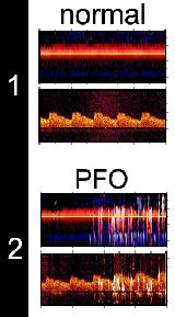
Transcranial Doppler (TCD) is a new, non-invasive way to diagnose a PFO.  It is extremely accurate, pain-free, and takes about 15 minutes.

A small ultrasound probe is placed on the temple just above the ear.  Ultrasound waves locate the flow of blood in the arteries of the head (see figure 1)

An intravenous is placed in the arm and a saline solution (sterile salt water) is injected.  The saline solution is filled with tiny dissolved microbubbles.

If there is no PFO, all the microbubbles are filtered by the lungs and no change is seen in the tracing of the blood flow.

If there is a PFO, some of the microbubbles pass (unfiltered by the lungs) through the PFO and travel to the arteries in the head.  The ultrasound waves are strongly reflected by even the smallest bubbles and are detected by the probe.  They are displayed as sharp color lines on the tracing (see figure 2). 

The injection is repeated while the patient takes a deep breath and then exhales forcefully as these maneuvers may be required to "open" the PFO.

The results of the test are available immediately and are discussed with the patient before he/she goes home. A copy of the results are forwarded to the primary care physician the same day.

The risk of this test is negligible.  There is no risk in injecting this microbubble saline solution as the "bubbles" are microscopic and completely dissolved in solution.

To arrange PFO screening using Transcranial Doppler (TCD) contact us.


To see a clip of a positive Transcranial Doppler (TCD) study click on the movie link
Media
TCD movie
Transesophageal Echocardiogram (TEE)

A PFO may also be detected by imaging it directly.  To do so, an ultrasound probe is placed from the mouth to the back of the throat.  In this way, the PFO can be seen very easily.  This test is somewhat uncomfortable and a mild intravenous sedative is used to ensure patient comfort and cooperation. 

The heart action is viewed "live" and the atrial septum and PFO (if present) is visualized.  Figure 1 shows the right atrium (RA) and left atrium (LA).  The arrow points to the trapdoor of the patent foramen ovale (PFO).

Saline solution is injected and the bubbles are easily seen in the right atrium (RA).  The presence of a PFO is confirmed by seeing bubbles pass to the left atrium (LA) through the PFO as is shown in figure 2.    

As with Transcranial Doppler the injection is repeated while the patient takes a deep breath and then exhales forcefully as these maneuvers may be required to "open" the PFO.

The results of the transesophageal echocardiogram are also available immediately and are discussed with the patient before he/she goes home. A copy of the results are forwarded to the primary care physician the same day.


Other heart tests and PFO

Either Transcranial Doppler (TCD) or a Transesophgeal Echocardiogram (TEE) is required to diagnose a Patent Foramen Ovale.

A PFO cannot  be diagnosed by:

  • Listening to the heart with a stethoscope
  • Electrocardiogram (ECG or EKG)
  • Stress test
  • Bloodwork
  • Transthoracic echocardiogram (echocardiogram done through the chest wall)*

* true in most instances


Click www.pfodoctor.org to return to main page